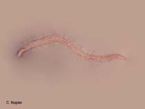
Polychaeta |
Amphinomida |
Amphinomidae
Environment: milieu / climate zone / depth range / distribution range
Ecology
Benthic; depth range 1 - 1100 m (Ref. 1456). Tropical
Indo-Pacific, Atlantic Ocean and the Mediterranean Sea. Tropical and subtropical.
Length at first maturity / Size / Weight / Age
Maturity: Lm ? range ? - ? cmmax. published weight: 12.54 g (Ref. 98347); max. published weight: 12.54 g
Minimum depth from Ref. 112705. Found on hard substrates (Ref. 112705), particularly under boulders (Ref. 3197), between tide marks (Ref. 78667), and in rock-pools (Ref. 89429). Sexual dimorphism: males are whitish because of the spermatozoa in the coelom; females are pinkish red due to the pinkish colored oocytes (Ref. 98347). In UAE, they are voracious carnivores feeding on sponges, hydroids, coral polyps and ascidians (Ref. 107862). Dwells in intertidal zones, in tidepools. Found in habitats dominated by Zoanthus pulchellus. Opportunistic carnivore (Ref. 132397).
Life cycle and mating behavior
Maturity | Reproduction | Spawning | Eggs | Fecundity | Larvae
Reproduces both asexually (regeneration) and sexually. It fragments along transverse fission planes called megasepta. The megaseptum involves an entire segment. The circular and longitudinal body wall muscles are partially lysed, and the gut is highly constricted. A megaseptum is dorsally transparent. The ventral pigment canals, however, are filled with brown granules on both sides of the megaseptum. The pigment canals are good reference points for the fission plane. These canals are epidermal channels just below the ventral and lateral longitudinal nerve cords and podial nerves. Fragmentation is accomplished by the constriction of longitudinal body wall fibers on both sides of the megaseptum. The latter is essentially inflexible. The body expands about the megaseptum and splits apart. The circular body wall fibers then contract to close the body cavity. The formation of a megaseptum requires one to two weeks. Fragmentation occurs within 10 to 15 minutes. In regeneration, the first setiger of an anterior fragment is reduced in size, and the parapodia are directed forwards. A translucent blastema is formed between the parapodia about 1 week after autotomization. This blastema enlarges and elongates. The first visible structures produced by the blastema are the prostomium, antennae, caruncle, eyes and palpi. The mouth and proboscis can not be seen. Segments appear to be produced within the first segment of the parental fragment. Incomplete intersegmental grooves are visible just in front of this segment. The parapodia are small and are similar to those of larger specimens. Some of the notosetae, however, are distally curved and truncate. They are also distally capped or hooded. Such notosetae seldom occur in adult specimens, and are generally absent from worms completing regenerative activities. The typical assortment of E. complanata setae are otherwise present. The branchiae resemble those of larger specimens, but are smaller. Similar events are observed in posterior regeneration. All regenerated tissues lack pigmentation, and external events can be easily followed. Anterior segments become pigmented soon after regeneration is completed. Pigmentation in posterior segments is a gradual process requiring many months. The ventral pigment canals become laden with brown particles. The pigment in these canals disappears soon after regeneration is completed. A conspicuous junction exists between the newly regenerated anterior segments and those of the original fragment. It persists after the segments have been replaced. These specimens were not included in the analyses of anterior regeneration rates. A similar junction is also present during the early phases of posterior regeneration, but soon disappears. During spawning, gametes are spawned through a pair of pygidial fold which are then forced through gut wall lesions into the intestine by constriction of body wall muscles. Intestinal cilia aid in the removal of gametes from the intestine. The pseudopore directs the gametes to either side of the body. Part of the intestine actually evaginates during this process and gives a false impression of entire pores. Males and females do not die after spawning. Larval development takes a total of 88 hours. Cleavage was typically spiral and holoblastic. Ciliated larvae were present 12 hours after fertilization. The posterior surface of the larva lacks ciliation at 32 hours. A pair of stiff hair like bristles is present. Two chaetoblast regions are visible at this stage. A trochophore larva is present after 48 hours. It has a transitory apical tuft. The prototroch is well defined. The first sign of the flotation setae can be seen at this stage. The membrane is deformed probably as a result of its penetration by the setae. The apical tuft is lost by 62 hours. The prototroch is more pronounced, and the floatation setae are three to five times longer than the larva. A metatrochophore is present after 72 hours. A telotroch is now present, as is a complete gut and a pair of eyespots. The larvae actively feed at this stage. Three setigers can be observed in larvae that are 88 hours old. The metatroch is located along the bottom margin of the anterior lobe. The telotroch surrounds the anus, additional trochal bands were not observed. The larvae begin to increase in size at this stage.
Gibbs, P.E. 1978 Macrofauna of the intertidal sand flats on low wooded islands, Northern Great Barrier Reef. Philosophical Transactions of the Royal Society of London, Series B 284(999):81-97. (Ref. 3197)
IUCN Red List Status
(Ref. 130435: Version 2025-1)
CITES status (Ref. 108899)
Not Evaluated
Not Evaluated
Threat to humans
Human uses
| FishSource |
Tools
More information
Population dynamicsGrowth
Max. ages / sizes
Length-weight rel.
Length-length rel.
Length-frequencies
Mass conversion
Abundance
PhysiologyOxygen consumption
Human RelatedStamps, coins, misc.
Internet sources
Estimates based on models
Preferred temperature
(Ref.
115969): 7.4 - 15.2, mean 10.1 (based on 611 cells).
Price category
Unknown.


